43 light microscope with labels
› microscopy › enZEISS Axioscope 5 Smart Laboratory Microscope Focus. Snap. Done. Forget about the 15 steps and clicks to document samples with multiple fluorescent labels. With Smart Microscopy, this is a thing of the past. Axioscope 5 with Axiocam 202 mono and Colibri 3 LED illumination take this workload from you. You keep your hands at the microscope stand. Relaxed. Light Microscope- Definition, Principle, Types, Parts, Labeled Diagram ... A light microscope is a biology laboratory instrument or tool, that uses visible light to detect and magnify very small objects and enlarge them. They use lenses to focus light on the specimen, magnifying it thus producing an image. The specimen is normally placed close to the microscopic lens.
› seterra › en-anMicroscope Components - Science Quiz - GeoGuessr Microscope Components - Science Quiz: The most common type of modern microscope is called a compound microscope. They have two systems of lenses, one is the eyepiece and the other is comprised of one or more objective lenses. This type of microscope has become so advanced that some are capable of magnifying up to 1000 times! Microscopes are used in almost all types of scientific research, and ...

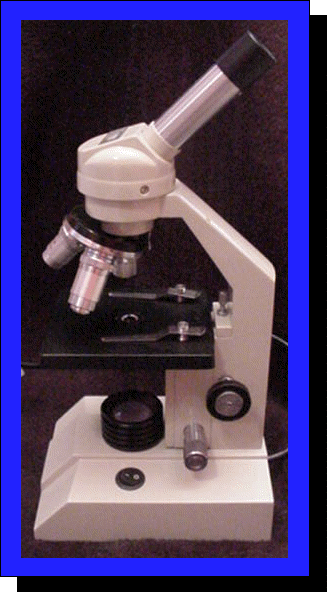
Light microscope with labels
Light Microscope Parts Labeled - 18 images - parts of the microscope ... [Light Microscope Parts Labeled] - 18 images - optical microscopy and specimen using the transmission, microscope with labels clip art at vector clip, solved microscope parts labeling 9 label the image of a c, , Electron microscope - Wikipedia An electron microscope is a microscope that uses a beam of accelerated electrons as a source of illumination. As the wavelength of an electron can be up to 100,000 times shorter than that of visible light photons, electron microscopes have a higher resolving power than light microscopes and can reveal the structure of smaller objects.. Electron microscopes use shaped magnetic … Label the Light Microscope - Labelled diagram - Wordwall Drag and drop the pins to their correct place on the image.. Eyepiece, Light Source, Base, Stage, Stage Clips, Fine Focus, Coarse Focus, Arm, Objective Lens.
Light microscope with labels. Microscope Labeling - The Biology Corner Microscope Labeling. This simple worksheet pairs with a lesson on the light microscope, where beginning biology students learn the parts of the light microscope and the steps needed to focus a slide under high power. The labeling worksheet could be used as a quiz or as part of direct instruction where students label the microscope as you go ... Compound Microscope Parts - Labeled Diagram and their Functions There are two major optical lens parts of a microscope: Eyepiece (10x) and Objective lenses (4x, 10x, 40x, 100x). Total magnification power is calculated by multiplying the magnification of the eyepiece and objective lens. The illuminator provides a source of light. The light is focused by the condenser and passing through the specimen placed ... Microscope Objective Lens | Products | Leica Microsystems Microscope Objectives. Leica Microsystems – The Ultimate in Optical Competence. For more than 170 years Leica Microsystems has designed and produced top-class objectives for a wide variety of applications in research, industry and medicine. The optics specialists at Leica Microsystems bring the highest level of experience and expertise to bear in reducing … Educational Atomic Force Microscope (AFM) - Thorlabs 05.11.2021 · The schematic to the right illustrates how the EDU-AFM1(/M) Educational AFM operates. Laser light is provided by a 635 nm fiber-coupled benchtop source. A patch cable feeds the laser light into a collimator consisting of a lens in an adjustable zoom housing.
Compound Microscope- Definition, Labeled Diagram, Principle, Parts, Uses The optical microscope often referred to as the light microscope, is a type of microscope that uses visible light and a system of lenses to magnify images of small subjects. The term "compound" in compound microscopes refers to the microscope having more than one lens. Devised with a system of combination of lenses, a compound microscope ... Microscope Labeling Game - PurposeGames.com About this Quiz. This is an online quiz called Microscope Labeling Game. There is a printable worksheet available for download here so you can take the quiz with pen and paper. This quiz has tags. Click on the tags below to find other quizzes on the same subject. Science. rsscience.com › stereo-microscopeParts of Stereo Microscope (Dissecting microscope) – labeled ... A stereo microscope allows you to see the surface of specimens with a 3-dimensional view. Under a stereo microscope, you can see the metallic texture and colors of the mosquito’s compound eyes. In contrast, the light has to pass through the specimen to form the image under a compound microscope. What is label in microscope? - Gowanusballroom.com Parts of the Microscope with Labeling (also Free Printouts) 1. Eyepiece. Through the eyepiece, you can visualize the object being studied. Its magnification capacity ranges between…. 2. Body tube/Head. It is the structure that connects the eyepiece to the lenses. Image 2: The body tube part of a….
Microscope, Microscope Parts, Labeled Diagram, and Functions Revolving Nosepiece or Turret: Turret is the part of the microscope that holds two or multiple objective lenses and helps to rotate objective lenses and also helps to easily change power. Objective Lenses: Three are 3 or 4 objective lenses on a microscope. The objective lenses almost always consist of 4x, 10x, 40x and 100x powers. The most common eyepiece lens is 10x and when it coupled with ... en.wikipedia.org › wiki › Electron_microscopeElectron microscope - Wikipedia An electron microscope is a microscope that uses a beam of accelerated electrons as a source of illumination. As the wavelength of an electron can be up to 100,000 times shorter than that of visible light photons, electron microscopes have a higher resolving power than light microscopes and can reveal the structure of smaller objects. Simple Microscope - Diagram (Parts labelled), Principle, Formula and Uses It is a type of optical microscope that uses visible light and lens to magnify objects. Despite the fact that they are rudimentary imaging devices, simple microscope finds use in microbiology to study biological specimens and microscopic organisms such as fungi, hydra and algae. They are also used by pedologists to study soil samples and ... Light microscope labels Flashcards | Quizlet Light microscope labels. STUDY. PLAY. Ocular lens. First automatic magnification (x10) Body tube. Holds ocular lense. Revolving nose piece. Holds and allows selection of desired objective lens. Lowest power objective lens. Veiws specimen at lowest megnification (x4) Medium power objective lens.
Histology Drawings: January 2014
Label the microscope — Science Learning Hub 08.06.2018 · All microscopes share features in common. In this interactive, you can label the different parts of a microscope. Use this with the Microscope parts activity to help students identify and label the main parts of a microscope and then describe their functions.. Drag and drop the text labels onto the microscope diagram. If you want to redo an answer, click on the …
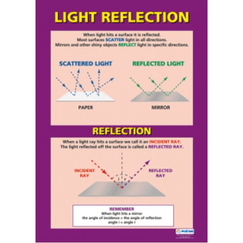
CHART, Light Reflection
Microscope Slide Labels - Histology and Light Microscopy Supplies ... Microscope Slide Labels: Pressure sensitive labels for microscope slides need no moistening. Self-sticking lables accept pencil, ball-point and felt-tip pen. 1000 labels per roll in convenient dispensing box.

Light Compound Microscope Labeled - Micropedia
proscitech.com.auProSciTech Laboratory supplies and Lab equipment for Histology, Pathology, Light Microscopy, Electron Microscopy and specialist researchers.

Types Of Light Microscopes
Microscope Types (with labeled diagrams) and Functions Is used to view samples that are not visible to the naked eye. Uses two types of lenses - Objective and ocular lenses. Has a higher level of magnification - Typically up to 2000x. Is used in hospitals and forensic labs by scientists, biologists and researchers to study micro organisms. Compound microscope labeled diagram.

Prokaryote Under Microscope
ZEISS Lightsheet 7 – Light Sheet Microscope Light sheet fluorescence microscopy (LSFM) is ideal for fast and gentle imaging of whole living model organisms, tissues and cells as they develop – over extended periods of time. What’s more, use ZEISS Lightsheet 7 to image large optically cleared specimens in toto – …
33 Label A Compound Light Microscope - Labels Database 2020
Parts of a microscope with functions and labeled diagram - Microbe Notes Q. Differentiate between a condenser and an Abbe condenser. Ans. Condensers are lenses that are used to collect and focus light from the illuminator into the specimen. They are found under the stage next to the diaphragm of the microscope. They play a major role in ensuring clear sharp images are produced with a high magnification of 400X and above.

high-definition light microscopes series for lab | ACOX
Microscope Parts and Functions Microscope Parts and Functions With Labeled Diagram and Functions How does a Compound Microscope Work?. Before exploring microscope parts and functions, you should probably understand that the compound light microscope is more complicated than just a microscope with more than one lens.. First, the purpose of a microscope is to magnify a small object or to magnify the fine details of a larger ...

compound light microscope labeled 28125 - Made By Creative Label
Parts of the Microscope with Labeling (also Free Printouts) 5. Knobs (fine and coarse) By adjusting the knob, you can adjust the focus of the microscope. The majority of the microscope models today have the knobs mounted on the same part of the device. Image 5: The circled parts of the microscope are the fine and coarse adjustment knobs. Picture Source: bp.blogspot.com.
Light Microscopes Educational-Line OBE | LabFriend Australia | Lab Equipment and Lab Supplies
Light Microscope Labeled - how scanning electron microscopes work ... Light Microscope Labeled - 16 images - senior biology cell theory microscopy, what is a light microscope with pictures, 29 you will love labeling a compound microscope db, microscope imaging station gallery,

Download And Use Microscope Png Clipart - Microscope Png , Free Transparent Clipart - ClipartKey
Compound Microscope Parts, Functions, and Labeled Diagram Compound Microscope Definitions for Labels. Eyepiece (ocular lens) with or without Pointer: The part that is looked through at the top of the compound microscope. Eyepieces typically have a magnification between 5x & 30x. Monocular or Binocular Head: Structural support that holds & connects the eyepieces to the objective lenses.

Review Questions
ZEISS Axioscope 5 Smart Laboratory Microscope Focus. Snap. Done. Forget about the 15 steps and clicks to document samples with multiple fluorescent labels. With Smart Microscopy, this is a thing of the past. Axioscope 5 with Axiocam 202 mono and Colibri 3 LED illumination take this workload from you. You keep your hands at the microscope stand. Relaxed.
34 Label Compound Light Microscope - Labels Database 2020
What is Electron Microscopy? - UMASS Medical School It is termed a scanning electron microscope because the image is formed by scanning a focused electron beam onto the surface of the specimen in a raster pattern. The interaction of the primary electron beam with the atoms near the surface causes the emission of particles at each point in the raster (e.g., low energy secondary electrons, high energy back scatter electrons, X-rays …
The Compound Light Microscope
Light Labs distributes PCR... Welcome to Light Labs. Since 2002, Light Labs has distributed high quality laboratory consumables and equipment, including MultiMax Barrier tips, PCR tubes and strip tubes, PCR plates, and much more. With an emphasis on customer service, we have successfully served the research marketplace with a wide array of laboratory goods.

Quia - 9AP Chapter 12 - The Cell Cycle (Detailed)
› products › microscopeMicroscope Objective Lens | Products | Leica Microsystems The objective lens is a critical part of the microscope optics. The microscope objective is positioned near the sample, specimen, or object being observed. It has a very important role in imaging, as it forms the first magnified image of the sample. The numerical aperture (NA) of the objective indicates its ability to gather light and largely determines the microscope’s resolution, the ...

Microscope World Blog: Bacteria under the Microscope
ProSciTech Laboratory supplies and Lab equipment for Histology, Pathology, Light Microscopy, Electron Microscopy and specialist researchers.

Diagrams of Microscope | 101 Diagrams
› microscopy › enZEISS Lightsheet 7 – Light Sheet Microscope This effect occurs in all fluorescence microscopes, but the illumination axis in light sheet fluorescence microscopy is perpendicular to the observation axis and so this effect is more obvious. In Lightsheet 7, a patented Pivot Scanner alters the angle of the light sheet upwards and downwards during image acquisition.

Microscope World Blog: Cheek Cells under Phase Contrast Microscope
Label the microscope — Science Learning Hub Label the microscope. Use this interactive to identify and label the main parts of a microscope. Drag and drop the text labels onto the microscope diagram. All microscopes share features in common. In this interactive, you can label the different parts of a microscope. Use this with the Microscope parts activity to help students identify and ...

New Microscope Reveals Stunning Detail | ZNZ Newsletter
Compound Microscope: Definition, Diagram, Parts, Uses, Working ... - BYJUS A compound microscope is defined as. A microscope with a high resolution and uses two sets of lenses providing a 2-dimensional image of the sample. The term compound refers to the usage of more than one lens in the microscope. Also, the compound microscope is one of the types of optical microscopes. The other type of optical microscope is a ...
Post a Comment for "43 light microscope with labels"