40 microscope images with labels
Label the microscope — Science Learning Hub Use this interactive to identify and label the main parts of a microscope. Drag and drop the text labels onto the microscope diagram. eye piece lens diaphragm or iris coarse focus adjustment stage base fine focus adjustment light source high-power objective Download Exercise Tweet › microscopy › enZEISS LSM 900 with Airyscan 2 - Compact Confocal Microscope ... Images were acquired with LSM 900 on ZEISS Celldiscoverer 7 using confocal GaAsP detectors (top row) and Airyscan 2 in HS mode (bottom row). Confocal images with LSM Plus (top, right) enhancing SNR and improving resolution of mitochondrial structures.
en.wikipedia.org › wiki › Scanning_electron_microscopeScanning electron microscope - Wikipedia A scanning electron microscope (SEM) is a type of electron microscope that produces images of a sample by scanning the surface with a focused beam of electrons.The electrons interact with atoms in the sample, producing various signals that contain information about the surface topography and composition of the sample.
Microscope images with labels
Microscope Labeling - The Biology Corner The google slides shown below have the same microscope image with the labels for students to copy. I often spend the first day walking students through the steps and having them look at a single slide as we do the steps. Students are often very enthusiastic about using microscopes and will try to start with the high power objective. Microscope Labeled Pictures, Images and Stock Photos Browse 49 microscope labeled stock photos and images available, or start a new search to explore more stock photos and images. Newest results Fluorescent Imaging immunofluorescence of cancer cells growing... Microscope diagram vector illustration. Labeled zoom instrument... Microscope diagram vector illustration. Parts of a microscope with functions and labeled diagram - Microbe Notes Optical parts of a microscope and their functions The optical parts of the microscope are used to view, magnify, and produce an image from a specimen placed on a slide. These parts include: Eyepiece - also known as the ocular. This is the part used to look through the microscope. Its found at the top of the microscope.
Microscope images with labels. Compound Microscope Parts - Labeled Diagram and their Functions The eyepiece (or ocular lens) is the lens part at the top of a microscope that the viewer looks through. The standard eyepiece has a magnification of 10x. You may exchange with an optional eyepiece ranging from 5x - 30x. [In this figure] The structure inside an eyepiece. The current design of the eyepiece is no longer a single convex lens. › NATIONAL-GEOGRAPHIC-Dual-StudentNational Geographic Dual LED Student Microscope Aug 07, 2017 · Buy NATIONAL GEOGRAPHIC Dual LED Student Microscope - 50+ pc Science Kit with 10 Prepared Biological & 10 Blank Slides, Lab Shrimp Experiment, Perfect for School Laboratory, Homeschool & Home Education: Microscopes - Amazon.com FREE DELIVERY possible on eligible purchases Microscope Labeling Game - PurposeGames.com About this Quiz. This is an online quiz called Microscope Labeling Game. There is a printable worksheet available for download here so you can take the quiz with pen and paper. This quiz has tags. Click on the tags below to find other quizzes on the same subject. Science. Mitosis Images Labeled | Virtual Anatomy Lab VAL - ncccval Endocrine Rabbit Dissection Unlabeled. Cardiovascular. Cardiovascular Histology Labeled. Cardiovascular Histology Unlabeled. Cardiovascular Models Labeled. Cardiovascular Models Unlabeled. Cardiovascular Sheep Heart Dissect-L. Cardiovascular Sheep Heart Disect-U. Cardiovascular Cat Dissection Labeled.
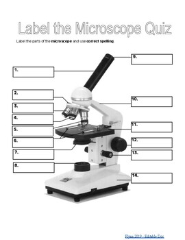
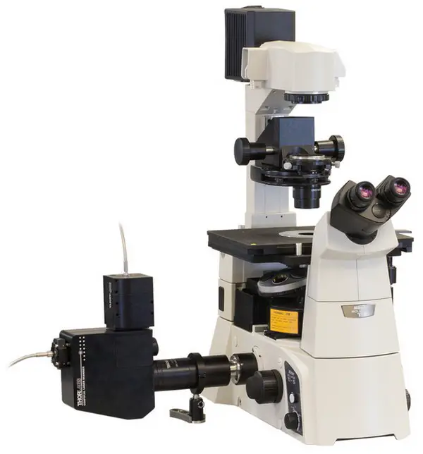
Microscope Label Interactive Worksheets & Teaching Resources | TpT 12. $1.89. PDF. Students will complete a timeline of the history of the microscope, label a diagram, and create a pocket foldable with terms and definition cards. The timeline can be completed according to the teacher's directions or like the answer key example. Optional cut & paste images and a QR code are a. Microscope Parts, Function, & Labeled Diagram - slidingmotion Microscope parts labeled diagram gives us all the information about its parts and their position in the microscope. Microscope Parts Labeled Diagram The principle of the Microscope gives you an exact reason to use it. It works on the 3 principles. Magnification Resolving Power Numerical Aperture. Parts of Microscope Head Base Arm Eyepiece Lens Simple Microscope - Parts, Functions, Diagram and Labelling Confocal microscope - It uses laser light to scan a dyed sample. Scanning electron microscope - Instead of light, this type of microscope uses electron. This type of microscope is used by researchers in the field of physical, biological, and medical science. Transmission electron microscope - it uses electron to create a magnified image ... › Avery-Self-Adhesive-RemovableAmazon.com: Avery Self-Adhesive Removable Labels, 0.75 x 1 ... Marking the expiration dates on white background labels like these are great. We purchased two sizes of the small Avery Labels and they have performed very well. Avery Removable Rectangular Labels, 0.5 x 0.75 Inches, White, Pack of 525 (6737) These are small size labels that we use for marking various products with expiration dates.
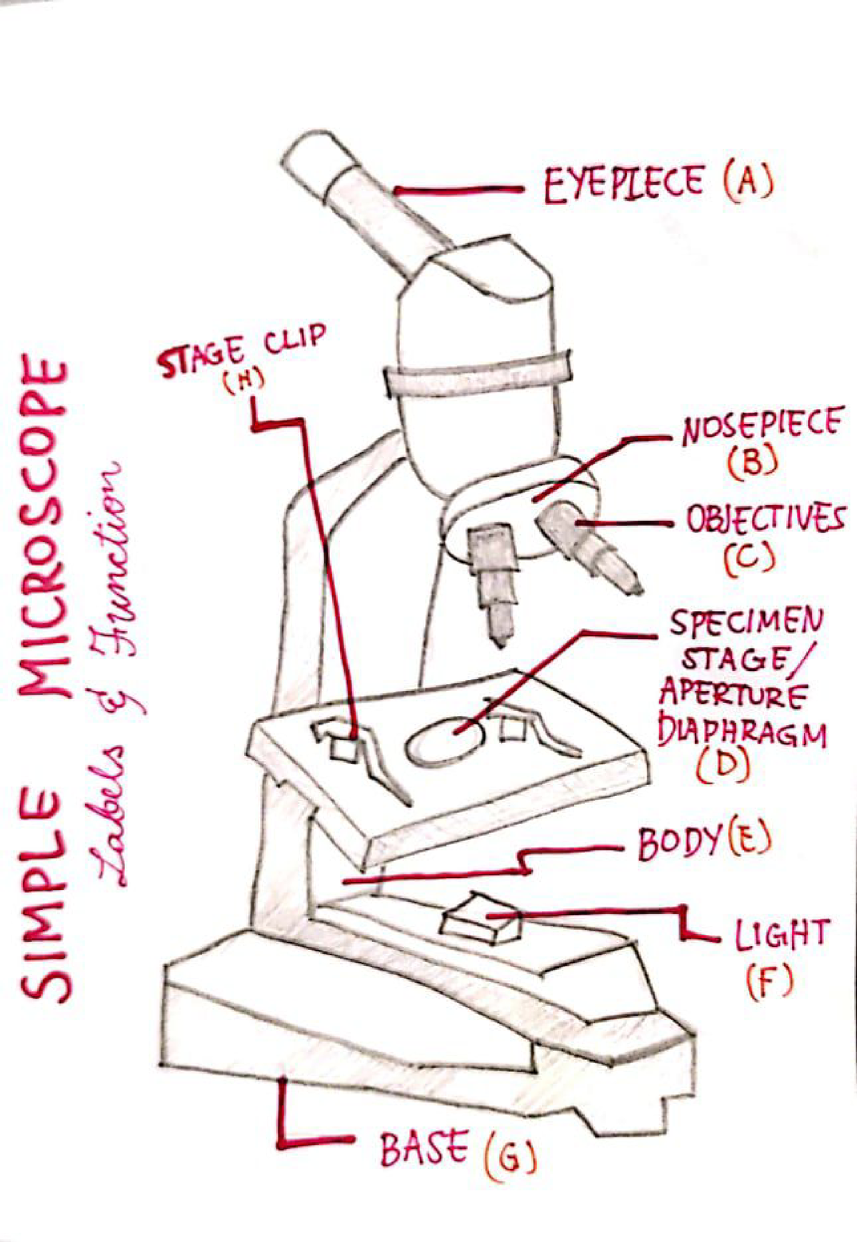
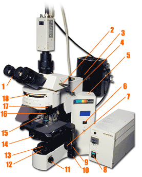
Microscope, Microscope Parts, Labeled Diagram, and Functions Revolving Nosepiece or Turret: Turret is the part of the microscope that holds two or multiple objective lenses and helps to rotate objective lenses and also helps to easily change power. Objective Lenses: Three are 3 or 4 objective lenses on a microscope. The objective lenses almost always consist of 4x, 10x, 40x and 100x powers. The most common eyepiece lens is 10x and when it coupled with ... A Study of the Microscope and its Functions With a Labeled Diagram ... The camera present within the microscope captures images to reveal the finer details of the specimen. This microscope can zoom and view the density of a specimen until it is only a micrometer thick and has a magnification ranging between 1,000 - 250,000x on the fluorescent screen. This microscope needs a computer software to yield precise ... Microscope With Labels clip art | Microscope parts, Scientific method ... clker.com vector clip art online, royalty free & public domain Download Clker's Microscope With Labels clip art and related images now. Multiple sizes and related images are all free on Clker.com. D Dixie Tsutsaeva 2k followers More information Microscope With Labels clip art Find this Pin and more on Art Journal Inspiration by Dixie Tsutsaeva. Parts of the Microscope with Labeling (also Free Printouts) Home Microscopes Parts of the Microscope with Labeling (also Free Printouts) By Editorial Team March 7, 2022 A microscope is one of the invaluable tools in the laboratory setting. It is used to observe things that cannot be seen by the naked eye. Table of Contents 1. Eyepiece 2. Body tube/Head 3. Turret/Nose piece 4. Objective lenses 5.
Microscope Labeling
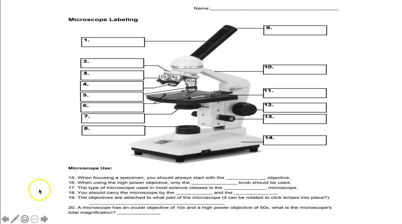
Labeling the Parts of the Microscope | Microscope World Resources Labeling the Parts of the Microscope This activity has been designed for use in homes and schools. Each microscope layout (both blank and the version with answers) are available as PDF downloads. You can view a more in-depth review of each part of the microscope here. Download the Label the Parts of the Microscope PDF printable version here.

Microscope Labeling Diagram | Quizlet
474,373 Microscope Images, Stock Photos & Vectors | Shutterstock Find Microscope stock images in HD and millions of other royalty-free stock photos, illustrations and vectors in the Shutterstock collection. Thousands of new, high-quality pictures added every day.
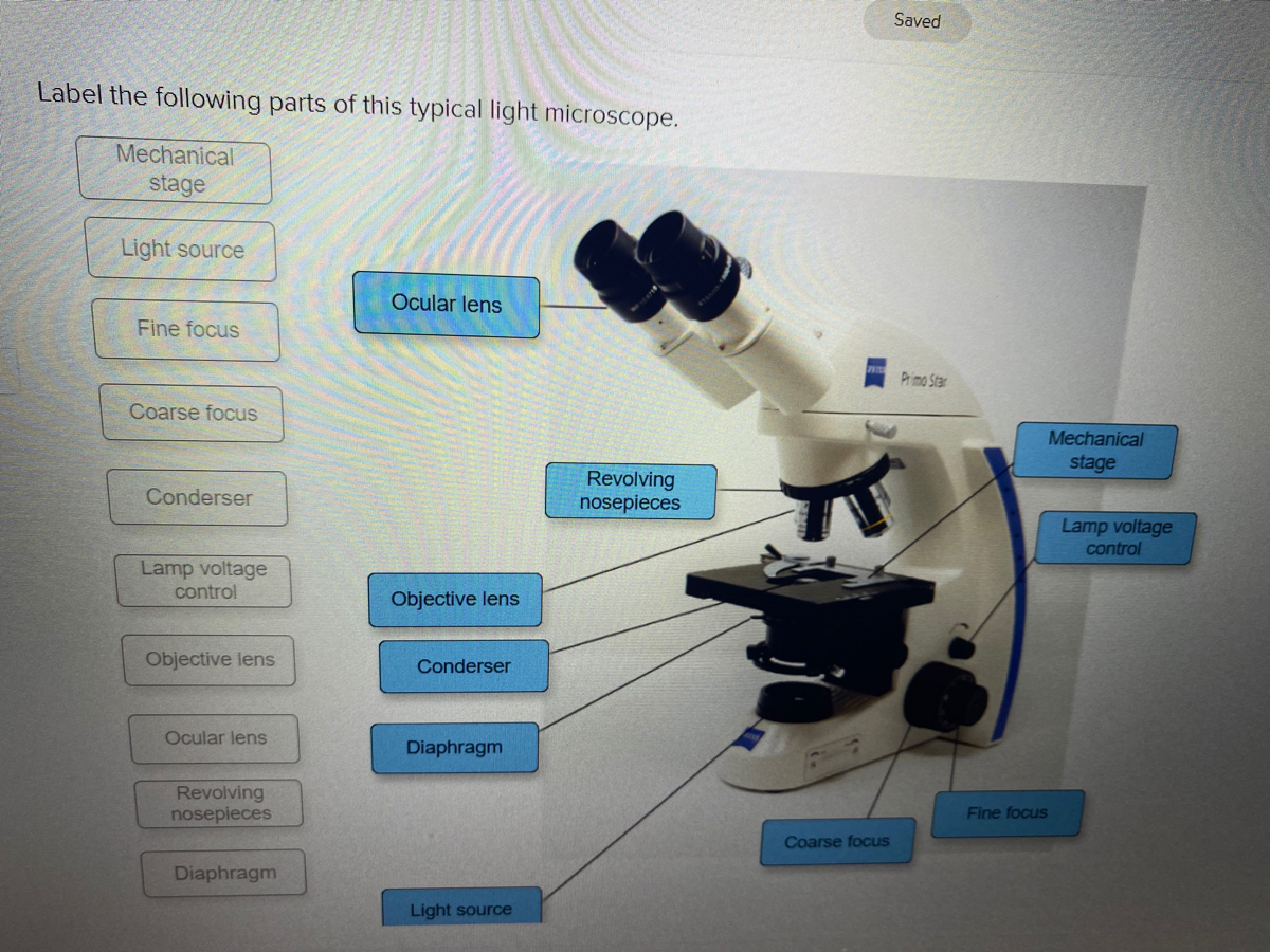
Answered: Saved Label the following parts of this… | bartleby
Microscope Types (with labeled diagrams) and Functions The working principle of a simple microscope is that when a lens is held close to the eye, a virtual, magnified and erect image of a specimen is formed at the least possible distance from which a human eye can discern objects clearly. Simple microscope labeled diagram Simple microscope functions It is used in industrial applications like:

Simple Microscope - Diagram (Parts labelled), Principle ...
18,701 Microscope drawing Images, Stock Photos & Vectors - Shutterstock Microscope drawing royalty-free images 18,701 microscope drawing stock photos, vectors, and illustrations are available royalty-free. See microscope drawing stock video clips Image type Orientation Color People Artists Sort by Popular Science Abstract Designs and Shapes College and University Art Styles Printing, Typography, and Calligraphy
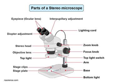
Parts of Stereo Microscope (Dissecting microscope) – labeled ...
› createJoin LiveJournal Password requirements: 6 to 30 characters long; ASCII characters only (characters found on a standard US keyboard); must contain at least 4 different symbols;
Parts of the Microscope Labeling Activity!
› microscopy › enZEISS Axioscan 7 Microscope Slide Scanner Digitize your specimens with Axioscan 7 – the reliable, reproducible way to create high-quality virtual microscope slides. Axioscan 7 combines qualities that you would not expect to get in a slide scanner: high speed digitization and outstanding image quality plus an unrivaled variety of imaging modes are all available in a fully automated and easy to operate system.

Free Microscope Drawing, Download Free Microscope Drawing png ...
22 Parts Of a Microscope With Their Function And Labeled Diagram Microscope Description A microscope is a laboratory instrument used to examine objects that are too small to be seen by the naked eye. In other words, it enlarges images of small objects. Invented by a Dutch spectacle maker in the late 16th century, light microscopes use lenses and light to magnify images. Generally a microscope ... Read more 22 Parts Of a Microscope With Their Function And ...
Microscope- Simple-AND Compound-WITH- Label - BS in Education ...
Microscope Parts and Functions Microscope Parts and Functions With Labeled Diagram and Functions How does a Compound Microscope Work?. Before exploring microscope parts and functions, you should probably understand that the compound light microscope is more complicated than just a microscope with more than one lens.. First, the purpose of a microscope is to magnify a small object or to magnify the fine details of a larger ...

Photo Compound microscope with labels Image #3850568
Electron Microscopy Images - Dartmouth We have a library of images recorded using our scanning and transmission electron microscopes. Some are shown below and others elsewhere. These images are in the public domain. If you have questions about the images or want some specific images contact Max Guinel . Hibiscus Flower (August 2021) Morphy Amorphophallus titanum anther cross section.
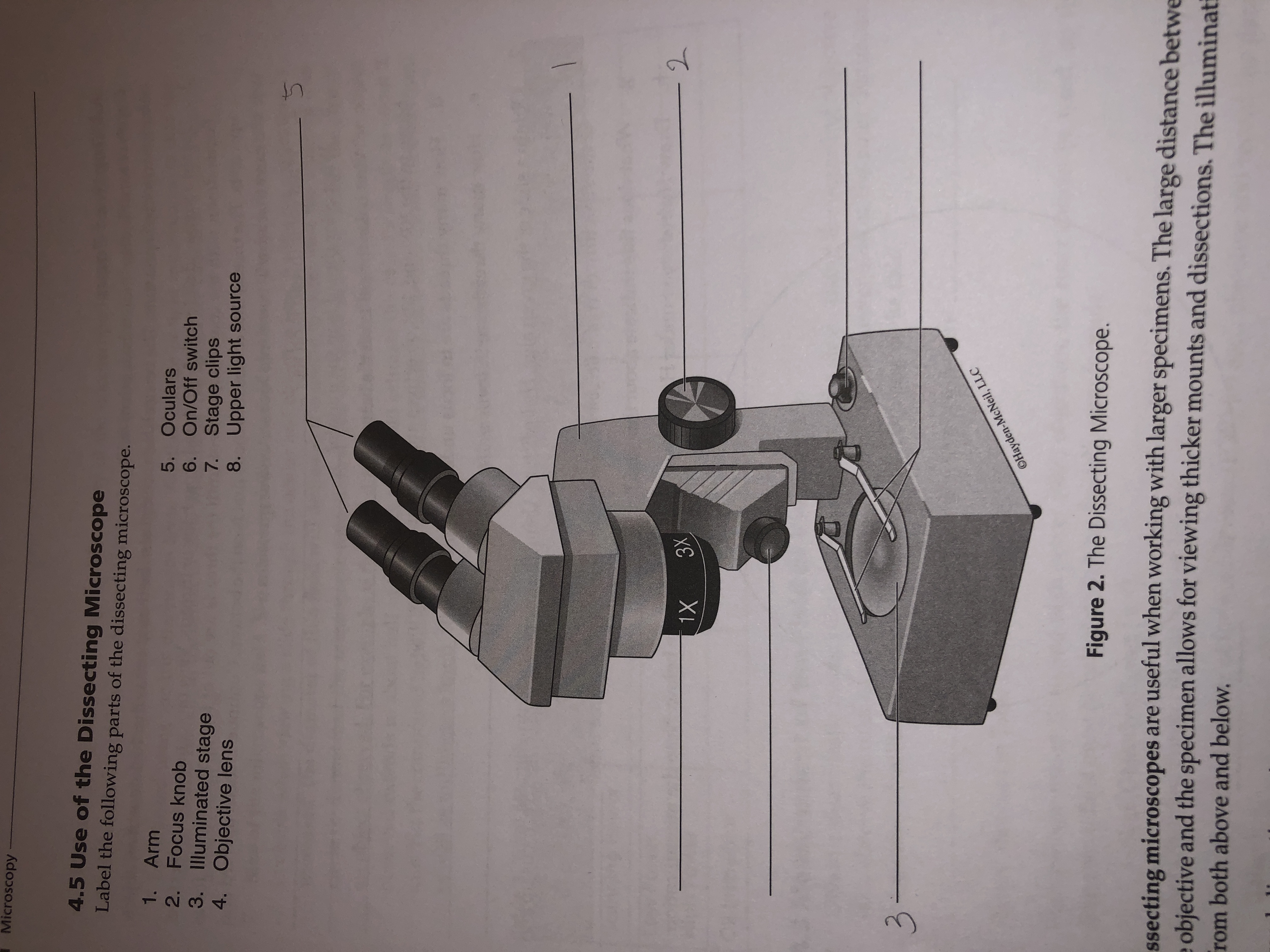
Answered: Microscopy 4.5 Use of the Dissecting… | bartleby
PDF Label parts of the Microscope Label parts of the Microscope: . Created Date: 20150715115425Z
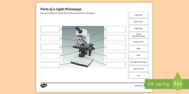
Parts of a Light Microscope Activity | Labeling Task
Amazing 27 Things Under The Microscope With Diagrams - Microbe Notes Figure: Hair under the microscope. Image Source: Microscope World. Observation under the stereo microscope. Stereo microscopes allow up to 90X magnification for the observation of the general structure and condition of the hair. The external characteristics like color, shape, texture, and length of hair can be seen easily through a ...

This is a common compound microscope. Label its parts from A ...
Microscope picture label Flashcards | Quizlet Start studying Microscope picture label. Learn vocabulary, terms, and more with flashcards, games, and other study tools.
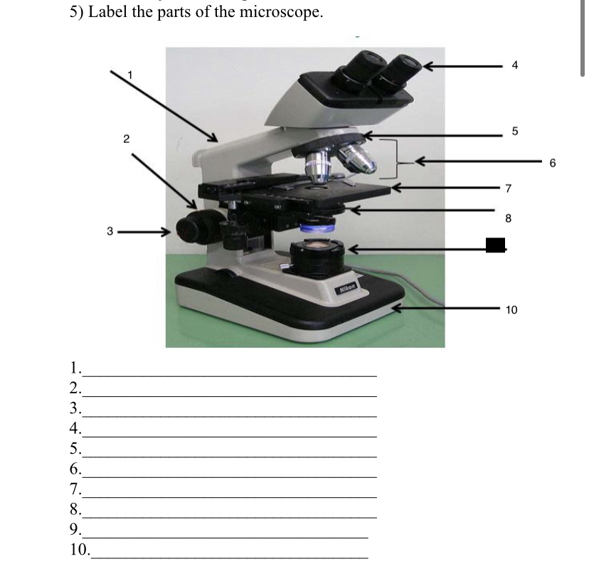
Answered: 5) Label the parts of the microscope. 1… | bartleby
A Study of the Microscope and its Functions With a Labeled Diagram ... May 21, 2019 - To better understand the structure and function of a microscope, we need to take a look at the labeled microscope diagrams of the compound and electron microscope. These diagrams clearly explain the functioning of the microscopes along with their respective parts.

Microscopes for Sale: Compound, Digital & Stereo | NY ...
Compound Microscope - Diagram (Parts labelled), Principle and Uses Image : Labeled Diagram of compound microscope parts See: Labeled Diagram showing differences between compound and simple microscope parts Structural Components The three structural components include 1. Head This is the upper part of the microscope that houses the optical parts 2. Arm

Microscope Labeling Activity - SMART Board Activity - Interactive Review
Explanation and Labelled Images - New York Microscope Company Fluorescence microscopy uses a high-intensity light source that excites a fluorescent molecule called a fluorophore in the sample observed. The samples are labeled with fluorophore where they absorb the high-intensity light from the source and emit a lower energy light of longer wavelength.

Microscope Diagram Labeled, Unlabeled and Blank | Parts of a ...
Compound Microscope Parts, Functions, and Labeled Diagram Compound Microscope Definitions for Labels Eyepiece (ocular lens) with or without Pointer: The part that is looked through at the top of the compound microscope. Eyepieces typically have a magnification between 5x & 30x. Monocular or Binocular Head: Structural support that holds & connects the eyepieces to the objective lenses.

Microscope labeled diagram
en.wikipedia.org › wiki › Electron_microscopeElectron microscope - Wikipedia An electron microscope is a microscope that uses a beam of accelerated electrons as a source of illumination. As the wavelength of an electron can be up to 100,000 times shorter than that of visible light photons , electron microscopes have a higher resolving power than light microscopes and can reveal the structure of smaller objects.
microscope with labels - Openclipart
400+ Free Microscope & Bacteria Images - Pixabay 412 Free images of Microscope Related Images: bacteria laboratory science scientist research biology lab virus microscopic Find your perfect microscope image. Free pictures to download and use in your next project.

Below is a photo of a compound light microscope with labels ...
Parts of a microscope with functions and labeled diagram - Microbe Notes Optical parts of a microscope and their functions The optical parts of the microscope are used to view, magnify, and produce an image from a specimen placed on a slide. These parts include: Eyepiece - also known as the ocular. This is the part used to look through the microscope. Its found at the top of the microscope.

Virtually Labeling a Microscope by Grace Voit | Teachers Pay ...
Microscope Labeled Pictures, Images and Stock Photos Browse 49 microscope labeled stock photos and images available, or start a new search to explore more stock photos and images. Newest results Fluorescent Imaging immunofluorescence of cancer cells growing... Microscope diagram vector illustration. Labeled zoom instrument... Microscope diagram vector illustration.

Compound Light Microscope Labeling Diagram | Quizlet
Microscope Labeling - The Biology Corner The google slides shown below have the same microscope image with the labels for students to copy. I often spend the first day walking students through the steps and having them look at a single slide as we do the steps. Students are often very enthusiastic about using microscopes and will try to start with the high power objective.
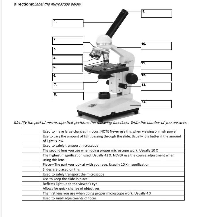
Solved Directions:Label the microscope below. 9. 1. 111 2 ...

National Ecoline D-ELDB Binocular Digital Microscope
Comparing and Contrasting the Different Parts of the Microscope

FUNRUI Kids Microscope, 450x, 200x, 100x Magnification Children Science Microscope Kit with LED Lights Includes Accessory Toy Set for Beginners Early ...
Lab - Microscope: MAH-Summer 2019-Anatomy and Physiology I

label the parts of microscope scope - Brainly.in

Meiji MT6500 Series PCM NIOSH 7400 Asbestos Microscope

Parts of a Microscope with Their Functions – Microbe Online

Biology label part of microscope

Biology Lab Quiz #2 (Labeling a Microscope) Diagram | Quizlet
Color the Microscope Parts

label microscope diagram | Charts | Microscope, Anatomy bones ...

Week 1: Microscope Usage & Snowflake Preservation
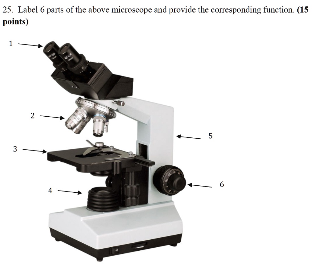
SOLVED: 25. Label 6 parts of the above microscope and provide ...

Microscope Diagram Labeled, Unlabeled and Blank | Parts of a ...
Getting Started - Virtual Fluorescent Microscope - Wartburg ...
Simple Microscope - Parts, Functions, Diagram and Labelling ...

Microscope Label Diagram | Quizlet

Label the light microscope | Teaching Resources
Post a Comment for "40 microscope images with labels"